Diagnosing Brain Metastases

Brain metastases occur when cancer cells from a primary tumor elsewhere in the body travel to the brain, forming secondary tumors. This can happen through the bloodstream or lymphatic system.
Various types of primary cancers, such as lung, breast, colon, and melanoma, can lead to brain metastases.
Understanding brain metastases is crucial because they differ significantly from primary brain tumors. Primary brain tumors originate within the brain, while brain metastases spread from elsewhere in the body.
Receiving a cancer diagnosis is life-changing, bringing fear, uncertainty, and many questions. Hearing "metastatic brain cancer" can be even more overwhelming.
However, it's important to remember that a cancer diagnosis marks the beginning of a journey toward understanding, treatment, and support.
How Are Brain Metastases Diagnosed?
Diagnosing brain metastases typically involves several steps, medical professionals, and diagnostic tests. Let's explore this process in detail:
Medical Evaluation
The process often starts with a visit to your primary care physician (PCP) or a specialist. They will ask about your medical history, including any history of cancer, and any recent or unusual symptoms. This information is crucial for an accurate diagnosis.
Why should you have your surgery with Dr. Cohen?
Dr. Cohen
- 7,500+ specialized surgeries performed by your chosen surgeon
- More personalized care
- Extensive experience = higher success rate and quicker recovery times
Major Health Centers
- No control over choosing the surgeon caring for you
- One-size-fits-all care
- Less specialization
For more reasons, please click here.
Neurological Examination
If brain metastases are suspected, the patient will be referred to a neurologist or neurosurgeon for a comprehensive neurological examination.
This examination assesses reflexes, muscle strength, coordination, and other neurological functions to identify any abnormalities that may suggest brain involvement.
Imaging Studies
Imaging studies play a pivotal role in diagnosing brain metastases. The most common imaging modalities used will include:
- Lesion Detection: Imaging enables the visualization and identification of metastatic lesions within the brain, constituting the primary step in diagnosis and treatment planning.
- Characterization of Lesions: Imaging studies provide detailed information regarding lesion size, location, and multiplicity, thereby assisting healthcare providers in assessing the extent and neurological implications of the disease.
- Treatment Strategy Determination: Knowledge of lesion characteristics, such as size and location, directly informs treatment decisions, aiding healthcare professionals in selecting the most suitable therapeutic approach—whether surgery, radiation therapy, or systemic treatments.
- Progress Monitoring: Post-treatment, imaging serves as a pivotal tool for tracking treatment response and evaluating the stability or regression of brain metastases over time. This facilitates ongoing assessment and adaptation of the treatment strategy.
What Imaging Tests Are Used To Diagnose Brain Metastases?
Diagnosing brain metastases typically involves the use of imaging studies to visualize and confirm the presence of metastatic lesions within the brain. The primary imaging modalities used for diagnosing brain metastases are:
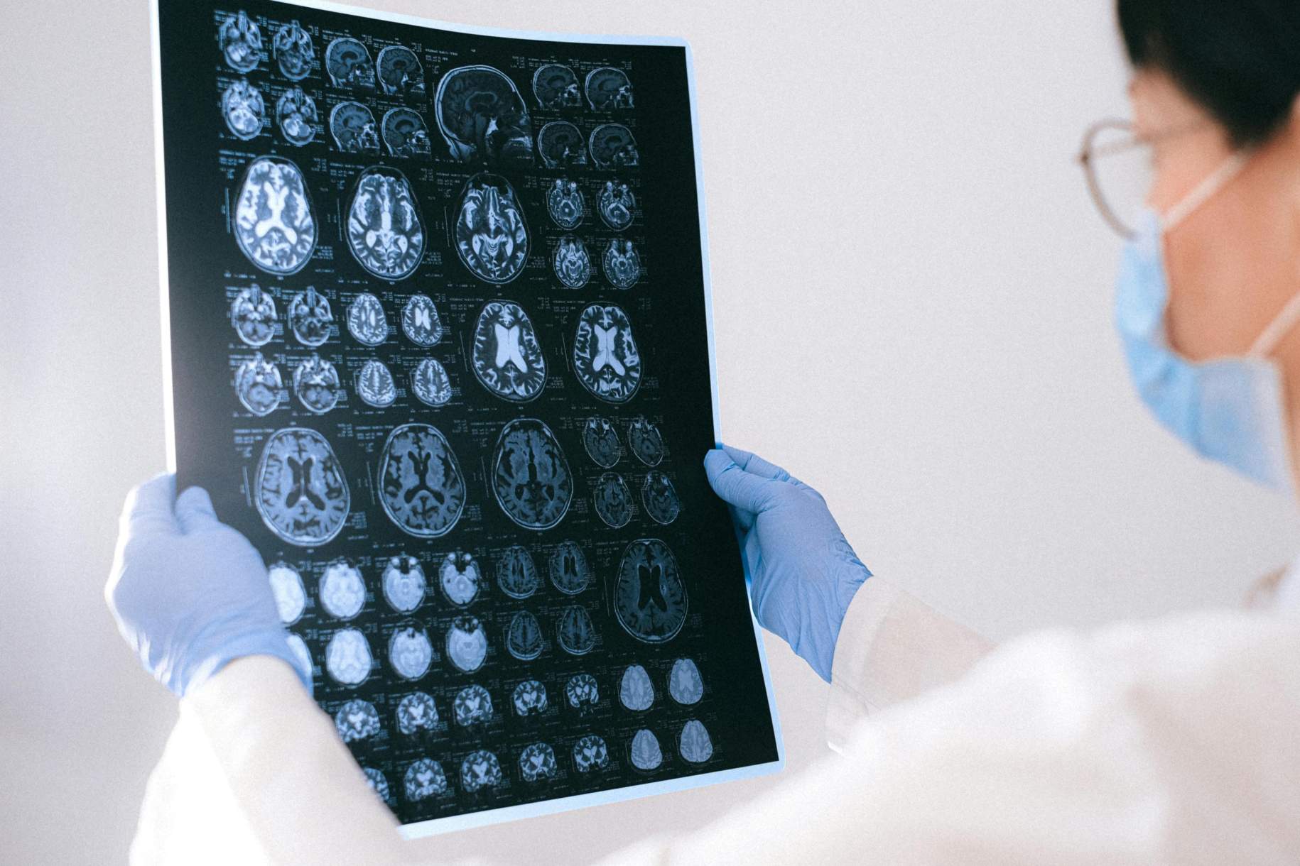
Magnetic Resonance Imaging (MRI)
MRI is considered the gold standard for detecting brain metastases. It provides highly detailed and multi-planar images of the brain.
This imaging technique allows for the visualization of tumors and provides crucial information about their size, location, and characteristics.
The use of contrast agents in MRI, such as gadolinium, enhances the visibility of lesions and aids in distinguishing them from normal brain tissue.
Computed Tomography (CT)
CT scans use X-rays and computer technology to create cross-sectional images of the brain.
While not as sensitive as MRI in detecting small lesions, CT scans are still valuable for assessing brain metastases.
CT scans are especially useful in situations where a rapid evaluation is necessary, as well as detecting calcified metastases, which can occur in specific cancer types.
In most cases, MRI is the preferred imaging modality due to its superior sensitivity and ability to provide detailed information about brain metastases. CT scans may be used when immediate assessment is required or when MRI is not immediately accessible.
Additionally, other specialized imaging techniques, such as Positron Emission Tomography (PET) scans and angiography (CTA or MRA), may be employed in specific clinical scenarios.
These studies are used to provide complementary information or to assess metabolic activity and blood flow within the brain. The choice of imaging modality and the specific approach to diagnosis will depend on individual patient factors, clinical symptoms, and the recommendations of the healthcare team.
Other Diagnostic Tools for Brain Metastases
Diagnostic tests that may be used to identify the primary cancer include:
- Blood tests: These can detect markers or abnormalities associated with certain types of cancer.
- Additional imaging studies: Scans of the chest, abdomen, and pelvis may be conducted to search for the primary tumor.
- Tissue biopsy: A tissue biopsy may be necessary to analyze cancer cells.
Identifying the primary cancer helps oncologists develop a comprehensive treatment plan that targets both the brain metastasis and the primary tumor.
The histopathological examination gives a detailed explanation of exactly what type of cells the tumor is made of and can help in differentiating a primary brain tumor from a metastatic brain tumor. It is also valuable in determining the further course of action concerning treatment strategies.
It is also a common scenario where brain metastases are discovered before the primary cancer is identified. Determining the primary cancer type is crucial for guiding treatment decisions and understanding the full extent of the disease.
Key Takeaways
- Brain metastases are usually diagnosed with a medical evaluation, neurological examination, and imaging tests.
- Imaging tests usually used are MRI, CT, and sometimes PET scans.
- The diagnosis of brain metastases is usually only confirmed by a microscopic histopathological examination of the brain.


