Diagnosing Hemangioblastoma

- Identifying the Symptoms and Where They Arise
- Imaging: The Window into the Brain and Spinal Cord
- Zooming In: Understanding What the Images Show
- Confirming the Diagnosis: Formal Angiography and Biopsy
- Supratentorial Hemangioblastoma: A Rare Variant
- Associated Risk Factors for Hemangioblastoma Diagnosis
- Your Medical Team: Guides on the Path to Diagnosis
- Key Takeaways
Hemangioblastomas are rare, benign vascular tumors that develop in the cerebellum, brainstem, or spinal cord. They are diagnosed through radiological imaging studies like magnetic resonance imaging (MRI) or computed tomography (CT) scans, though a definitive diagnosis is typically achieved through histopathological examination following surgical resection.
When faced with the possibility of a brain or spinal cord tumor, the path to diagnosis can seem daunting for patients and their families. With appropriate treatment and compassionate care, there is hope for managing symptoms and improving quality of life.
Identifying the Symptoms and Where They Arise
Hemangioblastomas are benign tumors that originate from the cells lining the blood vessels in the brain or spinal cord. Depending on where the tumor is located, symptoms can vary greatly.
A tumor in the brain may lead to headaches, vision problems, or difficulty with coordination. However, if it’s a spinal hemangioblastoma, it might cause back pain, weakness in the limbs, or problems with walking.
Seeking prompt medical attention if symptoms such as these appear is essential to achieving favorable outcomes and preserving neurological function.
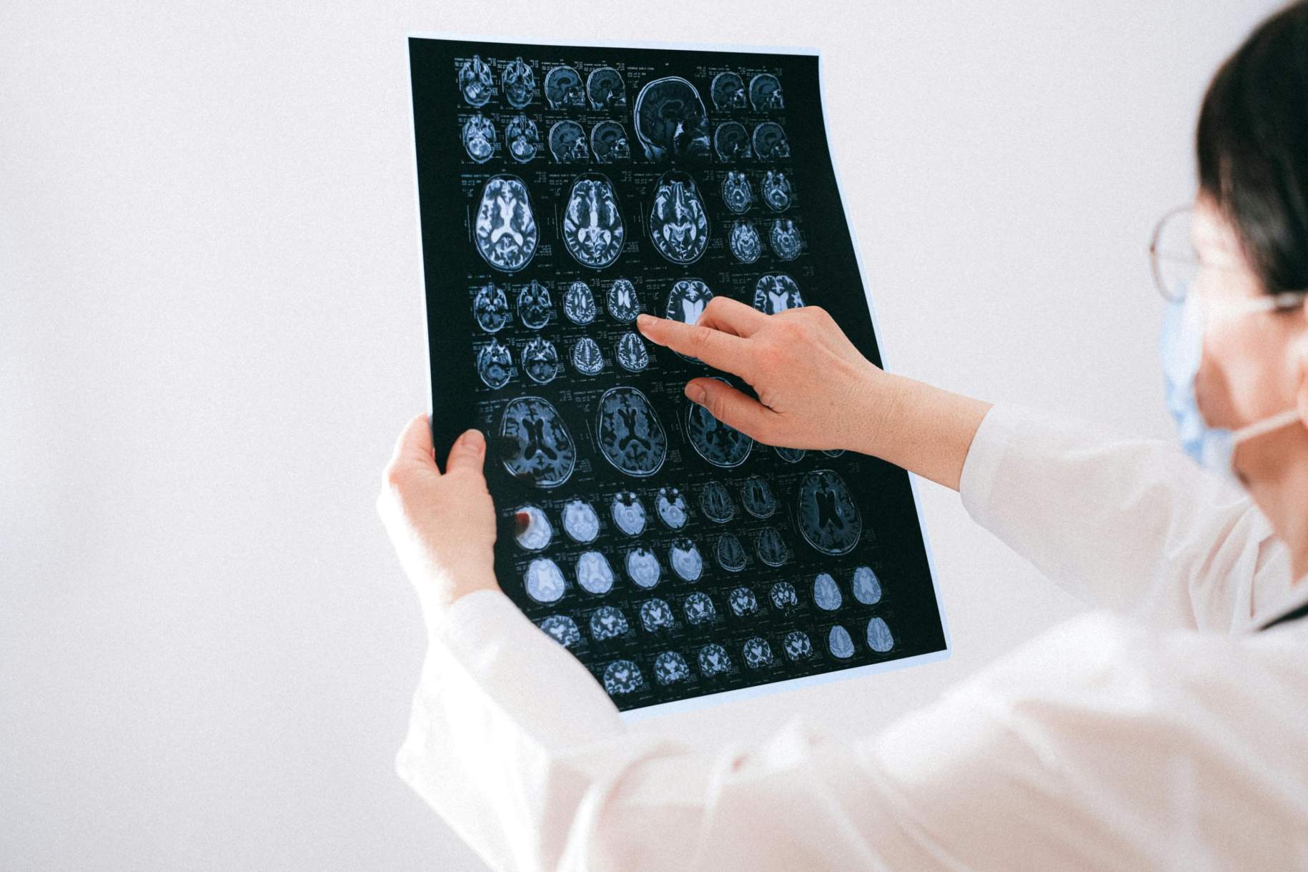
Imaging: The Window into the Brain and Spinal Cord
To visualize what's happening inside the brain or spinal cord, doctors rely on imaging tests like MRI and CT scans.
An MRI is a sophisticated imaging method that uses powerful magnets and radio waves to capture highly detailed images of the body’s internal structures. It's especially good at showing the soft tissues of the brain and can reveal the presence of a tumor.
A CT scan uses a series of X-rays to generate images of your bones and soft tissues. While these are faster than an MRI and can be used in urgent situations, they don’t show as much fine detail.
These imaging techniques play a critical role in diagnosing neurological tumors, offering crucial insights necessary to guide effective treatment decisions.
Why should you have your surgery with Dr. Cohen?
Dr. Cohen
- 7,500+ specialized surgeries performed by your chosen surgeon
- More personalized care
- Extensive experience = higher success rate and quicker recovery times
Major Health Centers
- No control over choosing the surgeon caring for you
- One-size-fits-all care
- Less specialization
For more reasons, please click here.
Zooming In: Understanding What the Images Show
When doctors review an MRI, they focus on identifying areas that stand out from normal brain or spinal cord tissue. Hemangioblastomas often appear as bright spots on the scan, especially with the use of a contrast dye.
Sometimes these tumors grow next to a cyst, which can look like a small water balloon on the scan. It is imperative that physicians can distinguish between different tumor types on these scans, as some types of brain tumors present in a similar manner.
For instance, pilocytic astrocytomas and medulloblastomas (other brain and spinal cord tumors) have some radiological similarities to hemangioblastomas. However, their unique features help physicians differentiate them.
Hemangioblastomas are highly vascularized with cysts, while pilocytic astrocytomas are also cystic but with less blood vessels. Medulloblastomas are solid, dense masses that do not have cystic components seen in other tumors.

Figure 1: MRI with contrast showing a hemangioblastoma located in the cerebellum.
Confirming the Diagnosis: Formal Angiography and Biopsy
While MRI and CT scans provide valuable insights, additional tests may be necessary to confirm a diagnosis.
One such procedure is forml angiography, which involves injecting a contrast dye into blood vessels of the brain and spinal cord to increase their visibility on X-ray images. This allows doctors to see the tumor’s blood supply and plan for potential surgery.
Another diagnostic tool is a biopsy, where a small tumor sample is taken and examined under a microscope. This is not performed routinely to aid in the diagnosis of hemangioblastoma, however, it may be used to ensure accurate diagnosis to guide the care team’s treatment plan.
Supratentorial Hemangioblastoma: A Rare Variant
Supratentorial hemangioblastomas are an exceedingly rare form of these tumors, accounting for only 2 to 10% of all cases. Unlike the more common cerebellar hemangioblastomas, these are found in the cerebral hemispheres, the brain’s largest region.
This region is responsible for executive functions like thought, memory, sensation, and voluntary movement. Despite their rarity, doctors use the same imaging tools, (MRI, CT, and formal angiography) to assess them.
These scans give doctors a clear picture of the size and location of the tumor, which helps them plan the best treatment. If you or a loved one is diagnosed with a supratentorial hemangioblastoma, your medical team will have personalized, compassionate conversations about what this means for treatment and prognosis.
Associated Risk Factors for Hemangioblastoma Diagnosis
While hemangioblastomas can occur spontaneously, certain risk factors increase the likelihood of diagnosis. Age is one such factor, with most cases occurring in adults aged 30 to 50.
Family history plays a significant role in hemangioblastomas associated with von Hippel-Lindau (VHL) disease. While there may be a genetic component, the majority occur in individuals with no family history of the condition.
In cases with no VHL association, other risk factors are less clear with most cases appearing sporadically. It's important for patients with a family history of brain tumors to discuss potential risks with their healthcare provider, as it may influence the need for surveillance or early diagnostic procedures.
Your Medical Team: Guides on the Path to Diagnosis
A dedicated group of healthcare professionals will work together to diagnose and treat a hemangioblastoma. This interdisciplinary team includes doctors who specialize in brain and spinal cord disorders, imaging experts who interpret scans, and surgeons who may perform procedures like tumor resections or biopsies.
Key Takeaways
- Hemangioblastomas are non-cancerous tumors that can cause a range of symptoms depending on their location in the brain or spinal cord.
- MRI is the most detailed imaging test for identifying hemangioblastomas. CT scans offer a quicker, less detailed view.
- Forma angiography and biopsy are procedures that can provide additional information to confirm a diagnosis.
- Genetic testing may be recommended if there's a family history of VHL disease or related tumors.


