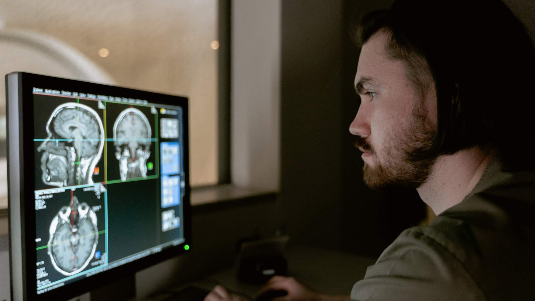
Diagnosing Meningioma


Meningiomas grow slowly and can be challenging to diagnose. Their symptoms may be mistaken for other conditions or written off as normal age-related changes. Depending on the location and size of the tumor, meningiomas can be completely asymptomatic or they can cause a variety of symptoms, including headaches, seizures, nausea and vomiting, weakness or numbness in the limbs, vision problems, and speech difficulties.
Herein, we will discuss some common physical examination and imaging findings used to diagnose meningioma.
Signs and Symptoms of Meningiomas
The slow-growing nature of a meningioma can prevent early detection. Patients may not experience any symptoms for years until the tumor becomes large enough to press on vital structures of the brain or spinal cord. Additionally, some patients with a meningioma do not experience any symptom at all—the tumor is only discovered by chance after medical imaging is performed for an unrelated condition. When symptoms do occur, they will depend on the size and location of the tumor as well as the presence of any edema (brain swelling) caused by the tumor. Common signs and symptoms for a meningioma may include:
- Headaches, usually dull or constant and worsen over time
- Seizures, especially with meningiomas located at the upper parts of the brain (supratentorial)
- Nausea and vomiting, especially in tumors located at the base of the skull or upper spine
- Trouble with vision, hearing, or speech if the tumor presses on nerves serving these functions
- Numbness or weakness in the arms or legs
- Confusion, drowsiness, or personality changes
How Are Meningiomas Diagnosed?
Diagnosing a meningioma can be challenging since most of these tumors are slow-growing and affect adults, who are more likely to attribute their symptoms to other conditions or even aging. There is no tell-tale or pathognomonic physical examination finding to definitively diagnose a meningioma.
As a result, meningiomas are often diagnosed using a combination of physical examination and imaging studies. In fact, it is more likely to identify a meningioma on routine imaging of the head for an unrelated condition than to recognize its obscure constellation of symptoms.
Why should you have your surgery with Dr. Cohen?
Dr. Cohen
- 7,500+ specialized surgeries performed by your chosen surgeon
- More personalized care
- Extensive experience = higher success rate and quicker recovery times
Major Health Centers
- No control over choosing the surgeon caring for you
- One-size-fits-all care
- Less specialization
For more reasons, please click here.
What Are the Common Findings in Patients With Meningioma?
Signs and symptoms of meningiomas vary by tumor location. While a physical examination alone cannot diagnose a meningioma definitively, it is important for your doctor to collect as many clues as possible during your consultation to rule in or rule out other conditions. During the consultation, your doctor will ask about your medical history and your presenting symptoms, including when they started, how often they occur, and if there are any triggers.
A neurological examination is imperative to check for abnormalities in your cranial nerves, motor skills, sensory abilities, reflexes, and balance. Any changes in your ability to think is also important to assess. If a brain tumor is suspected, radiological tests (e.g. CT, MRI) will be used to confirm the diagnosis.
Imaging Tests
Computed tomography (CT) and magnetic resonance imaging (MRI) are important diagnostic tools when evaluating a patient for a space occupying lesion, such as a brain tumor. Imaging studies focusing on the blood vessels within the head and neck (cerebral angiography) can also be used to identify a tumor’s blood supply.
CT Scans
CT imaging uses a series of X-rays to generate detailed images of the brain and is often one of the first imaging modalities obtained. Compared to MRI, CT is faster and cheaper. A CT scan suggestive of a meningioma contains an extra-axial (outside of the brain tissue) mass extending from the membranous covering of the brain (dural attachment). It is not uncommon for meningiomas to contain calcifications that also show up brightly on a CT scan. CT scans suggestive of an intracranial mass lesion (e.g., a tumor) are often followed up with an MRI that can provide higher resolution images of the brain.
MRI Scans
MRI uses a magnetic field and radio waves to create detailed images of soft tissue structures in the brain. An MRI scan can help evaluate the tumor's size, shape, and location and any nerves or other brain structures that may be affected by it. If you undergo surgery, MRI scans can also be used with neuronavigation technologies to guide the surgeon during the operation. Contrast dye, such as gadolinium, is often absorbed by tumors and is used during MRI to enhance visualization. Calcifications and a mass extending from the dural membrane (“dural tail”) are indicative of a meningioma.

Figure 1. MRI scans of a right frontal meningioma (large mass on the left side of each image) without contrast (left) or with contrast (right). In the right image, a bright white “dural tail” shows the tumor’s attachment to the outer membrane of the brain.
Biopsy
Meningiomas are reasonably well diagnosed on MRI but in very rare cases where imaging is not diagnostic and there are some questions about the aggressiveness of the tumor, a biopsy may be performed to remove a small tissue sample from the tumor for further evaluation under a microscope by a neuropathologist.
Certain cellular characteristics can help to determine if the meningioma is benign or aggressive. Biopsies are usually performed using a needle that is inserted into the brain through the skull. The biopsy sample is then sent to a laboratory for testing. Biopsies are not always necessary for meningiomas and should be discussed with your neurosurgeon.
Meningioma Grading
After diagnosis and during surgery, your doctor collects some samples of the tumor for biopsy analysis. These samples will be used to evaluate the grade of your meningioma. Meningioma grading is based on the physical appearance of tumor cells under a microscope. This helps to determine its severity and planning the best course of treatment. Meningiomas are typically graded according to the World Health Organization (WHO) into three grades: WHO Grade I (benign), WHO Grade II (atypical/borderline malignant), and WHO Grade III (malignant).
Low-grade meningiomas are slow growing, while high-grade meningiomas tend to be more aggressive and may invade brain tissue or even spread to other brain areas.
- WHO Grade I: Also called low-grade meningiomas, these tumors grow very slowly and are considered non-cancerous (benign.) They make up about 80% of meningiomas and are monitored with MRI scans every 6 to 12 months if undergoing observation and every year after surgery.
- WHO Grade II: These tumors grow more quickly than grade I and account for 18% of meningioma cases. Cells appear abnormal under a microscope with larger, more irregular shapes than those of grade I meningiomas. They are still considered borderline non-cancerous but have a higher recurrence rate than grade I tumors.
- WHO Grade III: Also called malignant meningiomas, these tumors are the most aggressive and make up 1%-3% of all meningioma cases. Grade III meningiomas contain cancerous cells and can proliferate and invade surrounding tissues. They are more likely to recur after treatment and may even spread to other brain and body parts.
Are Laboratory Tests Used to Diagnose Meningioma?
Blood tests can be used to look for inflammation, changes in protein levels, or other markers that could indicate the presence of cancer. However, many conditions can cause inflammation and alterations in normal protein levels so these cannot be used to definitively diagnose a meningioma. Lab tests mainly offer a baseline status of your general health and how other body organs are functioning.
What Other Conditions Present Similarly to Meningiomas?
In addition to meningioma, there are other health conditions that may present with similar symptoms. Some of the other conditions that may present similarly to meningiomas include:
- Other brain tumors such as gliomas and brain metastases
- Intracranial abscesses
- Seizures, migraines, and other neurological conditions
- Autoimmune disorders like multiple sclerosis
While these other conditions can present similarly to meningiomas, they often have different treatment options and prognoses. Therefore, it is crucial to seek a diagnosis from a qualified medical professional to ensure that you receive the best possible care and treatment.
Key Takeaways
Diagnosing meningiomas can be a complex process due to the slow-growing nature of the tumors and the overlap of symptoms with other conditions. To diagnose meningiomas, doctors often use a combination of physical examination findings and imaging tests. Treatment for meningiomas is dependent on many factors such as the location, size and symptoms associated with the tumor. Tumor grading also play a role in prognosis and management.


