Pineoblastoma: What the Patient Needs to Know

Pineoblastoma, also known as pinealoblastoma, is a rare cancerous tumor arising from the pineal gland. In this article we provide an overview of this condition, including its symptoms and treatment options.
What Is Pineoblastoma?
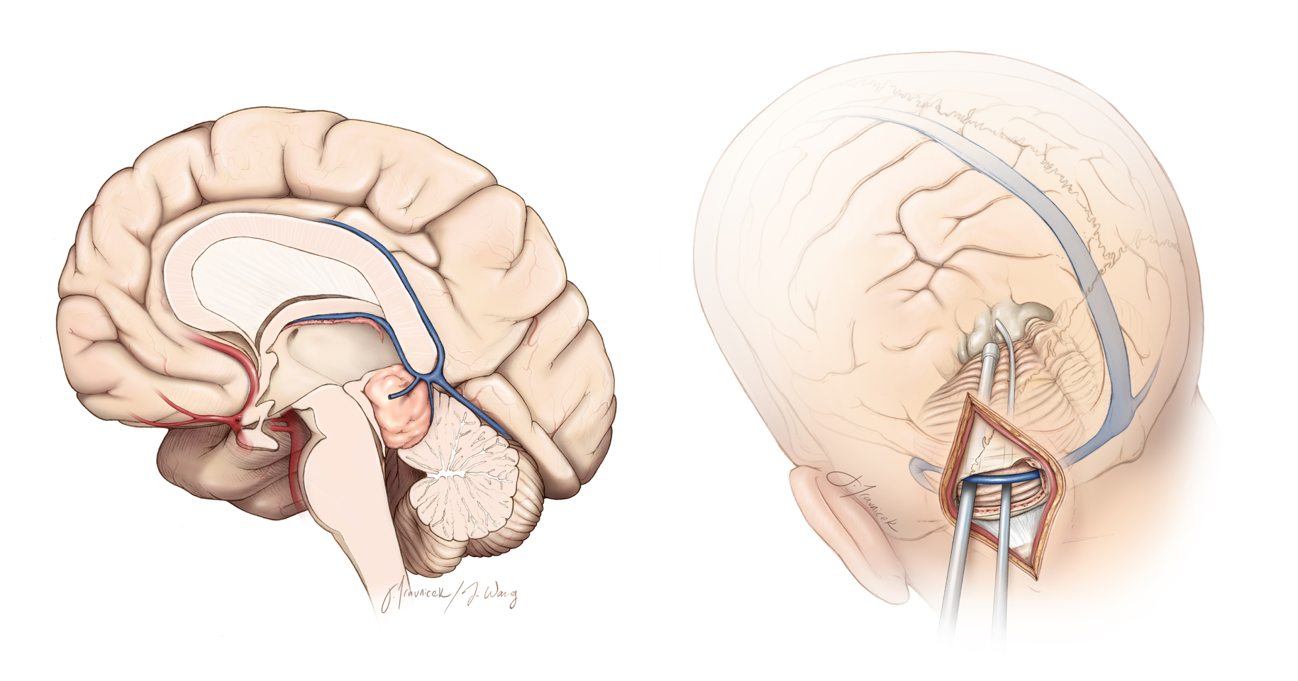
Pineoblastoma is an extremely rare and aggressive tumor type that originates from the pineal gland. The pineal gland is a small, pinecone-shaped structure located deep within the brain, between the two cerebral hemispheres. It is responsible for producing melatonin, a hormone that regulates sleep-wake cycles.
When a pineoblastoma forms, it can compress the surrounding nerves, parts of the brainstem, and ventricular system pathways. This can lead to a variety of symptoms. In many cases, blockage of the ventricular system can lead to increased pressure buildup in the brain in a life-threatening condition known as hydrocephalus.
What Are the Symptoms?
The most common presentation of pineoblastoma is that of nausea and vomiting combined with a change in cognitive function, behavior, or consciousness. This is most often due to buildup of pressure in the brain (hydrocephalus) caused by a large pineoblastoma obstructing the flow of cerebrospinal fluid.
Other symptoms can include the following:
- Headaches
- Difficulty with upward gaze, abnormal pupil reactions, and eyelid retraction (Parinaud syndrome)
- Problems with balance and coordination
- Sleep disturbances
- Vision problems, such as double vision or blurred vision
Why should you have your surgery with Dr. Cohen?
Dr. Cohen
- 7,500+ specialized surgeries performed by your chosen surgeon
- More personalized care
- Extensive experience = higher success rate and quicker recovery times
Major Health Centers
- No control over choosing the surgeon caring for you
- One-size-fits-all care
- Less specialization
For more reasons, please click here.
What Are the Causes?
The exact cause of pineoblastoma is not fully understood. Like many brain tumors, pineoblastomas are believed to result from genetic mutations that lead to abnormal cell growth and tumor formation. These mutations can be sporadic, meaning they occur by chance and are not inherited, or they can be part of a genetic syndrome.
One genetic condition associated with an incresaed risk of developing pineoblastoma is hereditary retinoblastoma, which is caused by mutations in the RB1 gene. This condition primarily affects the retina of the eye but can also increase the risk of pineal gland tumors.
How Common Is It?
Pineoblastoma is extremely rare and accounts for less than 0.1% of all brain tumors. Due to their rarity, large-scale studies on pineoblastoma are limited, and much of the knowledge about these tumors comes from case reports and small series.
The rarity of this tumor type also means that treatment protocols are often based on the collective experience of treating similar types of aggressive brain tumors. Thus, it is important to work with a supportive and experienced medical team.
How Is It Diagnosed?
Diagnosing pineoblastoma typically involves a combination of clinical evaluation, imaging studies, and sometimes tissue sampling. Here's how the process usually works:
1. Clinical Evaluation: A doctor will start by asking about symptoms and medical history. They'll also perform a physical examination, including a neurological exam to check for problems with balance, coordination, vision, and other brain functions.
2. Imaging Studies:
- MRI (Magnetic Resonance Imaging): An MRI scan of the brain is often the primary imaging study used to identify a pineoblastoma. It provides detailed images of the brain's structures and can show the location and size of the tumor.
- CT (Computed Tomography) Scan: A CT scan may be used if an MRI is not available or in emergency situations to quickly assess the brain.

Figure 1: MRI demonstrating a large abnormal growth in the pineal region.
3. Biopsy: If imaging studies show an abnormal growth within the pineal region, a biopsy may be performed to confirm the diagnosis. During a biopsy, a neurosurgeon removes a small sample of the tumor tissue, which is then examined under a microscope by a pathologist.
4. Additional Tests:
- Lumbar Puncture (Spinal Tap): This test may be done to check for tumor cells in the cerebrospinal fluid (CSF), which can indicate if the tumor has spread.
- Blood Tests: Certain blood tests can help rule out other conditions or assist in the diagnosis.
5. Staging and Assessment: If a pineoblastoma is diagnosed, additional imaging of the spine may be performed to determine if the tumor has spread, which is important for staging the disease and planning treatment.
In certain cases, patients may not be able to communicate their symptoms due to changes in their ability to think or speak. Imaging may demonstrate enlarged ventricles and hydrocephalus. Severe hydrocephalus is life-threatening and will require prompt intervention.
In these cases, a thin flexible catheter is placed into the ventricle at the bedside to drain CSF and relieve symptoms. This is called an external ventricular drain (EVD). The EVD is often a temporary measure that will be removed after surgery. In patients with persistent drainage problems, another procedure may be performed for permanent CSF diversion (ventriculoperitoneal shunt).
What Are the Treatment Options?
Surgery, radiation therapy, and chemotherapy are treatment options for pineoblastoma. Since pineoblastoma is exceedingly rare, there are no standardized treatment recommendations. The exact treatment plan will be tailored to your individual case.
Surgery may be performed first to obtain a tissue sample and remove the tumor during the same operation. If the tissue sample is suspicious for a pineoblastoma in the operating room, the surgeon can continue to remove as much of the tumor as is safely possible. Alternatively, a minimally invasive biopsy may be performed, with surgery occurring at a later date if necessary.
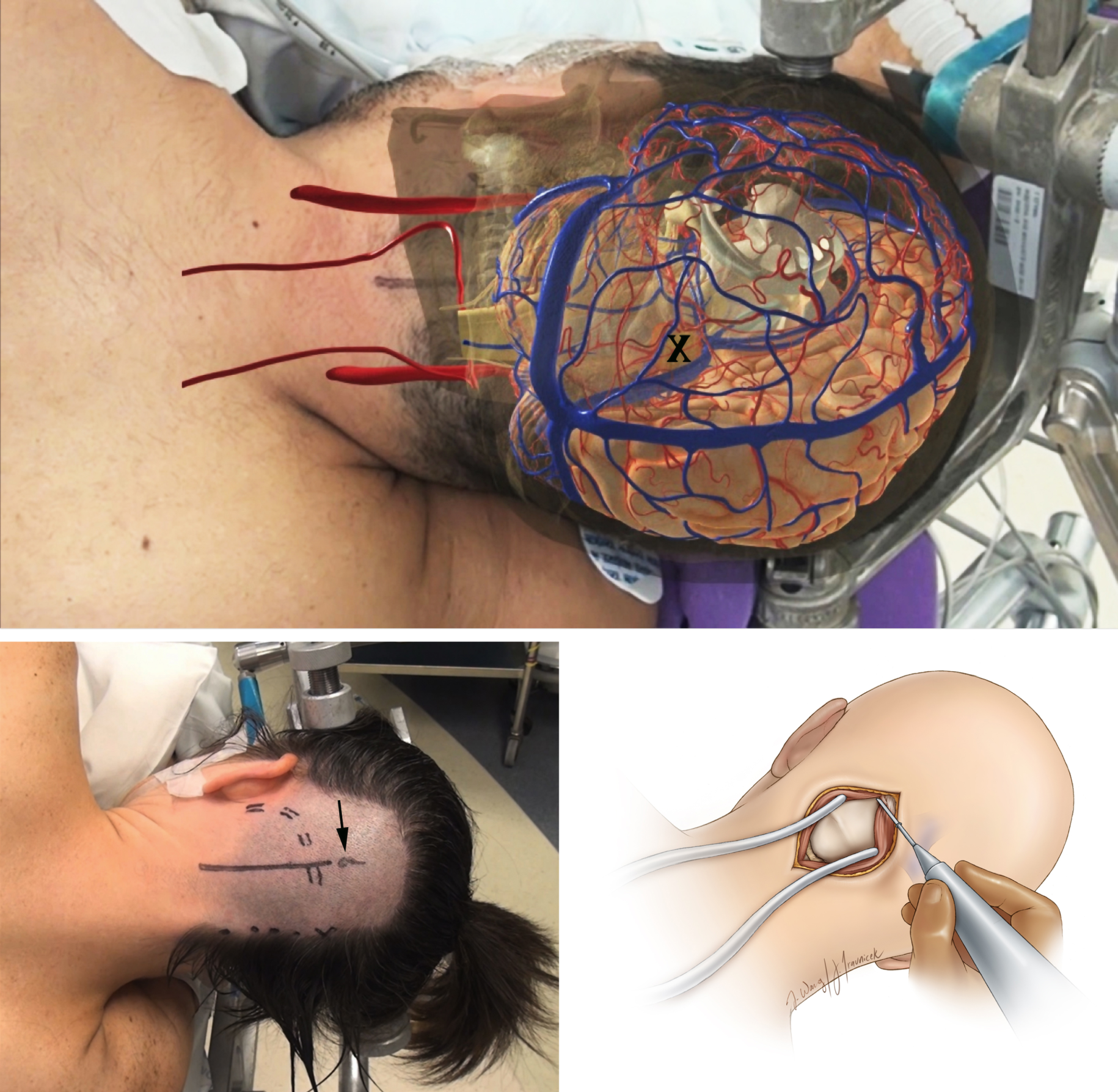
Figure 2: A patient is positioned for surgery. An overlay of the anatomical structures (top) helps the surgeon plan a path to the tumor. The incision path is marked on the scalp (bottom left). Once the scalp is incised, muscles are retracted to expose the skull bone (bottom right).
Radiation therapy is commonly administered after surgery to prevent growth from remnant tumor and tumor that has spread to distant sites. High-energy radiation beams are directed to the brain and spine (craniospinal radiation) with an extra dose delivered to the site of the tumor.
Chemotherapy is usually administered after radiation therapy. Common agents include vincristine and etoposide. Due to the damaging effects of craniospinal radiation on the developing brain, infants and young children often are not candidates for radiation therapy and may undergo chemotherapy alone.
What Is the Recovery Outlook?
With advances in therapy, the outlook for patients with pineoblastoma has improved significantly over the last few decades. However, it is still a challenging disease to effectively treat.
Since all pineoblastomas are considered grade 4 tumors, they are highly aggressive and prone to recur and spread. Approximately 15% of patients may have metastatic spread at the time of diagnosis. Maximal surgical removal is unlikely due to the tumor's ability to infiltrate the surrounding tissues and migrate to distant sites.
While radiation therapy and chemotherapy can help to prolong life, they cannot provide a complete cure for the disease. Reported survival times for pineoblastoma are extremely variable, with the proportion of patients alive at five years (5-year survival) ranging from 10% to over 80%.
Maintaining open communication with your medical team will be essential as you navigate through the journey ahead. Connecting with other families through patient foundations and forums can also provide valuable support and insight.
Key Takeaways
- Pineoblastoma is a rare malignant brain tumor that affects children more often than adults.
- Surgery, radiation, and chemotherapy are treatment options for pineoblastoma, but the exact regimen will depend on your individual case.
- The survival rates of pineoblastoma are highly variable, though in general are poor due to the aggressive nature of the disease.


