Radiosurgery for Arteriovenous Malformation

An arteriovenous malformation (AVM) is a tangle of abnormal blood vessel connections between arteries and veins that disrupt blood flow and distribution of oxygen to nearby tissues. Although surgery is typically curative, AVMs located in deep locations or neighboring critical structures may carry unacceptably high operative risks. In such cases, stereotactic radiosurgery may become a more favorable treatment option.
Understanding the risks and benefits of stereotactic radiosurgery will be important to determine whether this treatment option is right for you. Read on to learn more about stereotactic radiosurgery, how it works in the treatment for AVMs, and its associated risks.
What Is Stereotactic Radiosurgery?
Stereotactic radiosurgery is a type of radiation therapy that involves precise delivery of multiple radiation beams to a specified target location in the brain. These beams of radiation intersect at the center of the target to convey a high dose of radiation where the beams converge and a much lower dose to surrounding tissues receiving individual beams.
It can be helpful to understand what “stereotactic” and “radiosurgery” means to get a better sense of what this procedure entails. Stereotactic refers to image-guidance techniques that allow for accurate positioning of radiation beams to a target location within the body. This involves establishing a relationship between your surface anatomy with imaging tests (e.g., MRI) using various methods.
Radiosurgery, in contrast to its name, is not actually surgery. Instead, the inclusion of the word “surgery” is in reference to its highly precise nature and ability to target beams of radiation typically within less than a millimeter of tissue. The converged radiation beams are analogous to a scalpel or knife.
Stereotactic radiosurgery is a non-invasive treatment option that has been applied to destroy many types of intracranial lesions, such as brain tumors, that are inaccessible or inoperable via open surgery techniques.
In the case of AVMs, stereotactic radiosurgery provides a less invasive alternative to surgery. This can be favorable for AVMs that are deemed too risky to remove by open surgery. Stereotactic radiosurgery offers the highest chance of a cure for smaller AVMs less than 3 cm in diameter.
What Are the Different Types of Radiosurgery Systems?
Radiosurgery can be delivered by multiple systems as detailed below. The decision to choose one type of radiosurgery system over another may depend on what is most available or accessible to you in addition to the recommendations made by your neurosurgeon.
- Gamma rays (e.g., Gamma Knife): Focused gamma-rays (photons) are delivered to a specified target. Individual beams converge at the target location such that the highest radiation dose is provided to the volume of the lesion or the AVM.
- Linear accelerator-based systems (e.g., CyberKnife, Novalis): Focused x-rays (photons) are delivered to a specified target. Linear accelerator systems can move around the patient, allowing for more angles to approach the target, and more beams to divide the total radiation dose and therefore minimize radiation to surrounding tissues.
- Heavy charged particles (e.g., proton beam): Focused particles (e.g., protons) are delivered to a specified target. In comparison to photons (gamma and x-rays), protons deposit most of their energy at the target location and minimize radiation to adjacent areas. However, only a few proton beam therapy facilities are available around the world.
Why should you have your surgery with Dr. Cohen?
Dr. Cohen
- 7,500+ specialized surgeries performed by your chosen surgeon
- More personalized care
- Extensive experience = higher success rate and quicker recovery times
Major Health Centers
- No control over choosing the surgeon caring for you
- One-size-fits-all care
- Less specialization
For more reasons, please click here.
How Does Stereotactic Radiosurgery Work for AVMs?
Stereotactic radiosurgery can treat an AVM by damaging the walls of the blood vessels involved and causing it to scar and thicken over time. This eventually occludes the vessels feeding blood to the AVM, effectively removing an AVM from normal circulation.
Complete obliteration rates range from 50 – 90% depending on the size of the AVM. It is important to note that AVM exclusion via the use of radiosurgery occurs after a latency period of 2 to 3 years. Once complete obliteration is confirmed by an imaging test of the blood vessels (angiography), AVMs can be considered cured with a less than 1% risk of future hemorrhage.
Risks of Stereotactic Radiosurgery for AVMs
The main risks of stereotactic radiosurgery for AVMs are that of hemorrhage during the latency period, and damage to nearby brain tissues leading to neurological impairments. Stereotactic radiosurgery treatment will take several years until sufficient scarring occurs to obliterate the AVM. During this time, the risk of rupture remains.
Fewer than 10% of patients may experience adverse effects related to the radiosurgery procedure. Most are transient, but up to 3% of patients may suffer from permanent new neurological deficits. The type of impairment depends on the location of the AVM but can include the following:
- Headache
- Seizures
- Weakness or other movement disorders
- Sensory impairments
- Memory loss
- Vision problems
The risk of developing adverse effects appears to be higher when the AVM is in the brainstem or deep structures such as the thalamus. Delayed side effects such as brain tissue injury (radiation necrosis), brain swelling, and cyst formation may also occur in a small proportion of patients. These risks must be weighed against that of surgery when deciding which treatment option to proceed with.
What Does Stereotactic Radiosurgery for AVMs Entail?
Stereotactic radiosurgery is an outpatient procedure that involves several steps. Patients can typically go home the same day as the procedure. The general steps are detailed below.
Preparation
A box-like head frame is secured to the scalp and skull with special pins. The scalp is numbed while the pins secure the frame to the skull by penetrating the scalp and a few millimeters of skull bone. Patients may feel discomfort and pressure during this process. This frame is used for adequate immobilization during the procedure and to plan the trajectory of the radiation beams.
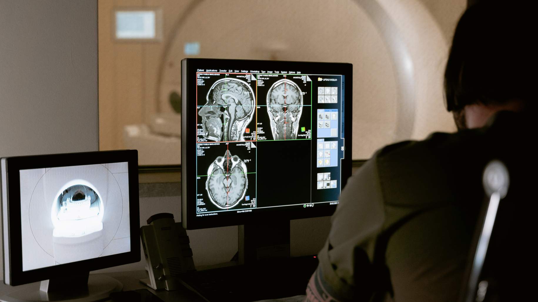
Imaging Tests
Once the frame is placed on the head, an MRI will be taken to visualize the location of intracranial structures. The MRI produces a 3D image of the brain which can be used in combination with the reference points on the frame to accurately target the AVM.
A cerebral angiogram is also performed by injecting a contrast dye into a blood vessel while taking x-ray images. This shows the specific blood vessels involved in the AVM which will be important when determining the boundaries of the radiation dose.
Planning
The MRI and angiogram are used to outline exactly where the radiation beams will target. Since the boundaries of an AVM are irregular, the treatment team must ensure that the radiation dose conforms exactly to the perimeter of the AVM. This planning step may take longer than the actual treatment time and span a few hours.
Treatment
The actual treatment session is painless and requires you to lie still in the radiosurgery machine for approximately 30 – 50 minutes. After the procedure, the frame is removed and patients typically go home the same day. The scalp may feel tender at the points where the pins were placed.
After radiosurgery, an MRI will be performed every 12 months or so until complete AVM obliteration is observed in 3 years via an angiogram. AVMs that are still present after several years, or those that are large may receive additional radiosurgery treatment sessions.
Key Takeaways
- Stereotactic radiosurgery is a non-invasive outpatient treatment option typically considered for small AVMs that are difficult or risky to surgically remove.
- Stereotactic radiosurgery can cure select AVMs in 50 – 90% of cases after a latency period of 2 to 3 years.
- Small AVMs, less than 3 cm in, size are more likely to be completely obliterated after stereotactic radiosurgery.


