What Is the Process for Diagnosing Glioma?

Written by: Aaron Cohen-Gadol, MD
Last Updated: October 4, 2024
See our Medical Editorial Standards for more information about how we maintain the highest standards of medical accuracy.
If you have symptoms of glioma, your primary physician may refer you to a neurologist or neurosurgeon for further testing. This article will provide insight into the symptoms, exams, and tests used to diagnose gliomas.
Please note that the some of the more common symptoms of any brain tumor can be nonspecific and overlap with the symptoms of many other conditions. Therefore, symptoms alone cannot diagnose a glioma or any other brain tumor. A thorough workup including dedicated imaging is necessary to diagnose a glioma in a timely fashion.
Diagnosing glioma begins with the physician examining and interviewing the patient. After the provider recognizes potential symptoms of a brain tumor, they direct their patients for further testing. These tests often include computed tomography (CT or CAT) scans, magnetic resonance imaging (MRI), and possibly a biopsy.
To provide an idea of what to expect, we will discuss these procedures in more depth. Also, we will answer some common questions. At what age are gliomas usually diagnosed? What is an MRI Scan? How does a biopsy work?
These are good questions. Below, we offer some guidance.
How Do Doctors Diagnose Glioma?
To diagnose a potential glioma, physicians will take a stepwise approach. As you will see, the procedures move from simple to more in-depth.
Doctors often use the following methods in this order to diagnose glioma:
- Neurological exam
- CT scan
- MRI scan
- Biopsy
Neurological Exam to Diagnose Glioma
A physical exam alone will not diagnose glioma. However, health providers will use the physical exam to guide further diagnostic decisions. For example, a physician might order more tests if a patient loses coordination, has repeated vomiting, or reports worsening headaches.
In addition to advanced neurological assessments of speech, reflexes, balance and strength, physicians may order blood tests to look for signs of other diseases or disorders which may be causing symptoms.
Why should you have your surgery with Dr. Cohen?
Dr. Cohen
- 7,000+ specialized surgeries performed by your chosen surgeon
- More personalized care
- Extensive experience = higher success rate and quicker recovery times
Major Health Centers
- No control over choosing the surgeon caring for you
- One-size-fits-all care
- Less specialization
For more reasons, please click here.
CT Scan for Diagnosing Glioma
Hospitals can perform CT scans quickly. The test usually only lasts a few minutes. The CT scan offers a fast look inside the brain. Claustrophobia is not an issue here as the scan is relatively open and comfortable for the patient to complete.
What is a CT scan? A CT scan uses similar technology to an X-ray; however, the CT scan sends beams through the body at different angles. Then, a computer takes these frames and configures them into a clear image. The CT scan is painless, and all the patient must do is lay on a bed while the machine performs the test.
CT scans are a common first step in diagnosing a glioma. However, physicians often move on to another, more advanced scan, like an MRI.
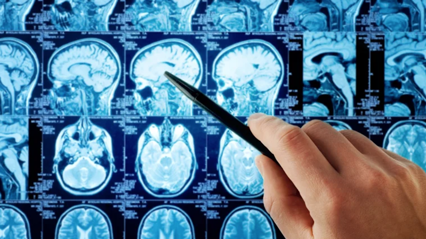
MRI to Diagnose Glioma
After looking at a CT scan, physicians may order an MRI to get a closer look at a potential glioma. The MRI test takes longer than a CT scan, lasting around 30 to 45 minutes. What is an MRI? MRI stands for magnetic resonance imaging. The MRI scanner uses magnets and radio waves to record the movement of protons in water molecules in your body. It is a fairly complex machine; however, it yields much clearer results than a CT scan.
Since the MRI scan takes place in a more closed space, claustrophobia can be an issue for certain patients. In these cases, the physician can prescribe a short acting sedative to help with the discomfort just for the purposes of completing the scan.
The MRI is beneficial in several ways. First, it provides a clear picture of soft tissue (the brain). Second, it carries less risk than a CT scan as MRI does not use ionizing radiation. The MRI is also painless, only requiring the patient to lay still.
One thing to know is that if you have any implanted devices (such as a pacemaker or defribillator), you may not qualify for an MRI. Some devices are approved for MRI and will be evaluated prior to undergoing imaging. Also, like the CT scan, the MRI might use contrast dye. If you have had allergic reactions to contrast dye, notify your physician.
Brain Tumor Biopsy Procedure
Once your physician has found a tumor and you have been referred to a neurosurgeon, the neurosurgeon may decide to perform a biopsy to finalize the diagnosis before making a more definitive decision for removal of the tumor through a craniotomy.
What is a biopsy? A biopsy is a surgical procedure under general anesthesia that uses a small needle or precise instrument to remove and collect a piece of the tumor for microscopic examination by the pathologist. As some brain tumors can look alike on MRI, further testing such as a biopsy is needed for final accurate diagnosis. Please refer to the stereotactic biopsy chapter for further details.
A biopsy can provide definitive information on the properties of the brain tumor, leading to more focused treatments. Moreover, some tumors are located within critical parts of the brain and are therefore not safely amenable to surgical removal. In these cases, the biopsy results will guide the next steps of radiation and/or chemotherapy based on the specific genetic makeup of the tumor.
What Is the Average Age of Glioblastoma Diagnosis?
Glioblastoma is the most aggressive form of brain cancer. The average age for diagnosis of glioblastoma is around 64 years old. However, that does not mean that other age groups are unaffected. Pediatric patients (children), aged 0-19, make up 6% of all brain tumors. Adolescents and young adults, aged 15-39, comprise 14.5% of all brain tumors.
Brain tumors do not discriminate by age; however, they are more common in older adults. Also, glioblastomas appear more common in males.
The Different Grades of Glioma
An important part of the diagnostic process involves determining the glioma grade. Brain tumors are unlike other types of cancer, they use unique grading system with four grades. These grades will describe the type of glioma and provide insight into their prognosis.
There are four grades of glioma:
- Grade I: These are relatively benign, and surgeons can usually remove them with good outcomes.
- Grade II: Grade II gliomas tend to grow into a higher grade. Often treated first with surgery.
- Grade III: These tumors are dangerous and carry the risk of growing into fully malignant grade IV.
- Grade IV: Glioblastomas fall into this category. Grade IV is the most cancerous grade, with the survival period often under two years.
Depending on the glioma grade, physicians might perform more diagnostic tests to improve their treatment decisions.
Key Takeaways
- Diagnosing a glioma can be a journey. Physicians will combine physical examination results with advanced procedures like an MRI and biopsy to make a definitive diagnosis.
- Unfortunately, many gliomas are diagnosed in their higher grades as an early diagnosis is not possible. This journey can be very anxiety provoking and affects the entire family. Well informed patients are best empowered to make the journey more tolerable.