Risk Factors and Causes of Craniopharyngioma


Craniopharyngiomas are tumors that develop near the pituitary gland and at the base of the brain. They are rare, with an incidence rate of 0.5 to 2 cases per 1 million people, and account for 1% to 3% of all brain tumors in children. They more commonly affect children aged 5 to 14 years and older adults aged 50 to 74 years.
Although craniopharyngiomas are generally benign tumors, they can cause serious health problems due to their location at the base of the brain. If you or someone you know is affected by a craniopharyngioma, you may be concerned about the risks of developing this tumor. This comprehensive guide will answer some of the most frequently asked questions to help patients and caregivers better understand this condition.
Craniopharyngioma Origin
Craniopharyngiomas originate from remnants of Rathke’s pouch, a small sac-like structure that is present in the developing fetus and later forms part of the pituitary gland by the time we are born. Normally, a small piece of Rathke’s pouch persists after birth. In rare cases, mutations in these cells can lead to the formation of a craniopharyngioma.
Because craniopharyngiomas arise from remnants of a structure that initially formed part of the pituitary gland, these tumors are typically located in close proximity to the pituitary gland and its surrounding structures. Besides the pituitary gland, surrounding structures of critical importance include the hypothalamus and optic nerves, which carry visual information from the eyes to the brain.
Craniopharyngioma Causes
Craniopharyngiomas are sporadic tumors, meaning that they occur randomly with no known cause. Thus, there are no definitive risk factors that have been identified as contributing to their development.
They exhibit no racial or gender bias and do not appear to be associated with any other medical conditions or genetic predispositions. There are, however, several symptoms that are commonly seen with a craniopharyngioma, depending on the location and size of the tumor. Common symptoms of craniopharyngioma include:
- Headache due to increased intracranial pressure
- Visual complications, such as blurred or double vision
- Stunted growth or delayed puberty in children
- Fatigue and low blood pressure
- Nausea and vomiting
- Personality changes and confusion
Why should you have your surgery with Dr. Cohen?
Dr. Cohen
- 7,500+ specialized surgeries performed by your chosen surgeon
- More personalized care
- Extensive experience = higher success rate and quicker recovery times
Major Health Centers
- No control over choosing the surgeon caring for you
- One-size-fits-all care
- Less specialization
For more reasons, please click here.
Craniopharyngioma Age
Craniopharyngiomas exhibit a bimodal age distribution by more commonly affecting children aged 5 to 14 years and older adults aged 50 to 74 years. In older adults, craniopharyngiomas are often associated with other medical conditions and may require various treatments to manage their symptoms.
How Can You Prevent Craniopharyngioma?
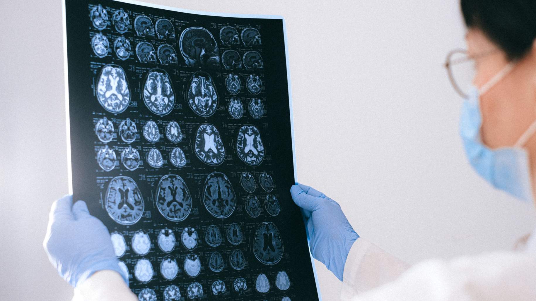
Currently, there is no known way to prevent the development of craniopharyngiomas. As such, it is important for patients to visit their primary care physician regularly to monitor for any symptoms. Diagnosis typically involves laboratory and imaging tests, such as MRI or CT scan, to confirm the presence of a craniopharyngioma and determine the best course of treatment.
Craniopharyngioma treatment is often determined by the size and location of the tumor and the age and health of the patient. Treatment options for craniopharyngiomas include surgery, radiotherapy, and chemotherapy.
Due to the complexity of treatment, patients and caregivers need to work closely with their medical and surgical teams, and discuss all treatment plans carefully. This is essential in ensuring the best possible outcome and successful recovery.
Craniopharyngioma Calcification
Calcification refers to the buildup of calcium deposits in the body's tissues. In craniopharyngiomas, calcification typically occurs in the cysts of the tumor and may be identified on diagnostic imaging such as an MRI or CT scan.
Although calcification is not an indication of malignancy, it can often be seen in larger tumors and may be associated with an increased risk of recurrence. Calcification is a common occurrence in craniopharyngioma, especially the adamantinomatous type.
Are You Born With Craniopharyngioma?
Craniopharyngioma is not a genetic condition, meaning it does not have any known inheritable factors or associations with congenital disorders. These tumors are derived from remnants of embryonic tissue in the brain and occur randomly with no known cause.
In extremely rare cases, babies may be born with a craniopharyngioma. This may occur due to mutations during fetal development that lead to abnormal cell growth and a tumor large enough to cause symptoms by the time of birth. However, older children aged 5 to 14 and adults over 50 are more commonly affected.
Is Craniopharyngioma Genetic?
Craniopharyngiomas are not thought to have a genetic cause, meaning they are not passed down from parents to children. Researchers suspect that tumor development is a random event that occurs during embryonic development (embryogenesis), with no known cause, resulting from metaplasia (abnormal cell transformation) of the craniopharyngeal duct and surrounding structures.
Although the effects of craniopharyngiomas during their early stages of development are mild, they can cause serious damage and complications if left untreated. As the tumors grow, they exert pressure on surrounding brain tissues and the pituitary gland. This can cause headaches, visual problems, hormonal imbalances, and cognitive impairment.
Key Takeaways
Craniopharyngioma is a rare and benign tumor that occurs near the pituitary gland and hypothalamus. It is most common in children aged 5 to 14 and adults over 50 and has no known cause. If large, it can compress surrounding structures such as the pituitary gland, hypothalamus, and optic nerves, and cause symptoms.


